---
author:
- contributor_roles: []
family: Edmunds
given: Scott
url: https://orcid.org/0000-0001-6444-1436
blog:
authors: null
community_id: 52db0518-e228-4260-8c54-c4e323b2569d
created: 1675555200
current_feed_url: null
description: Data driven blogging from the GigaScience editors
doi: https://doi.org/10.59350/gigablog
favicon: https://rogue-scholar.org/api/communities/52db0518-e228-4260-8c54-c4e323b2569d/logo
feed_format: application/atom+xml
feed_url: http://gigasciencejournal.com/blog/feed/atom/
filter: null
generator: Other
home_page_url: https://gigasciencejournal.com/blog
issn: null
language: eng
license: https://creativecommons.org/licenses/by/4.0/legalcode
prefix: '10.59350'
relative_url: null
secure: false
slug: gigablog
status: archived
subfield: '1311'
title: GigaBlog
updated: null
use_api: null
container: GigaBlog
date: '2015-11-17T00:00:00+00:00'
date_updated: '2025-12-06T10:34:47+00:00'
guid: http://blogs.biomedcentral.com/gigablog/?p=1615
identifier: https://doi.org/10.59350/3mykx-9wf86
image: https://s.w.org/images/core/emoji/14.0.0/72x72/1f600.png
images:
- height: '150'
src: http://gigasciencejournal.com/blog/wp-content/uploads/2015/11/brittlestar-150x150-1.jpg
width: '150'
- alt: "\U0001F600"
src: https://s.w.org/images/core/emoji/14.0.0/72x72/1f600.png
- alt: Jannes et al., AJMS 2015
height: '235'
sizes: '(max-width: 492px) 100vw, 492px'
src: http://gigasciencejournal.com/blog/wp-content/uploads/2015/11/Figure-2.jpeg
srcset: http://gigasciencejournal.com/blog/wp-content/uploads/2015/11/Figure-2.jpeg,
http://gigasciencejournal.com/blog/wp-content/uploads/2015/11/Figure-2-300x143.jpeg
width: '492'
- src: http://gigasciencejournal.com/blog/wp-content/uploads/2015/11/Figure-2.jpeg
issn: null
keywords:
- Biology
- Technology
- Africa
- Big Data
- GigaDB
lang: en
license: https://creativecommons.org/licenses/by/4.0/legalcode
reference:
- id: https://doi.org/10.1186/s13742-015-0093-2
unstructured: Landschoff, J., Du Plessis, A., & Griffiths, C. L. (2015). A dataset
describing brooding in three species of South African brittle stars, comprising
seven high-resolution, micro X-ray computed tomography scans. GigaScience,
4(1).
- id: https://doi.org/10.5524/100130
unstructured: Du Plessis, A., Landschoff, J., & Griffiths, C. L. (2015). 3-dimensional
microCT reconstructions of brooding brittle stars [Data set]. GigaScience
Database.
- id: https://doi.org/10.2989/1814232x.2015.1095801
unstructured: Landschoff, J., & Griffiths, C. (2015). Three-dimensional visualisation
of brooding behaviour in two distantly related brittle stars from South African
waters. African Journal of Marine Science, 37(4), 533–541.
- id: http://gigasciencejournal.com/blog/brooding-brittle-star-babies-qa-jannes-landschoff/
unstructured: Unknown title
- id: http://gigasciencejournal.com/blog
unstructured: Unknown title
rid: g7npe-2jk56
rights: https://creativecommons.org/licenses/by/4.0/legalcode
summary: '**Pregnant Starfish go Big Data. **Brittle stars are a class of organisms
that are closely related to starfish. While most members of this group reproduce
externally, there are some species that develop their young internally and ''give
birth'' to live young.'
title: Brooding Brittle Star Babies. Q&A with Jannes Landschoff
url: https://wayback.archive-it.org/22098/2025-05-01T17:13:42Z/http://gigasciencejournal.com/blog/brooding-brittle-star-babies-qa-jannes-landschoff
version: v1
---
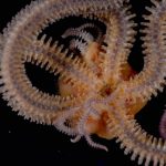
\*\*Pregnant Starfish go Big Data.\
\*\*Brittle stars are a class of organisms that are closely related to
starfish. While most members of this group reproduce externally, there
are some species that develop their young internally and \'give birth\'
to live young. While sonograms are the means by which humans can get a
look at their developing offspring, live-bearing brittle stars have not
been so lucky, as researchers primary means to investigate the brooding
chambers and juveniles of brittle stars has been via dissection. This
technique destroys the sample, thus eliminating the possibility of
further study and sharing of samples for others to use. In new work
[published in
*GigaScience*](https://doi.org/10.1186/s13742-015-0093-2){target="_blank"
rel="noopener noreferrer"} (supporting a [coordinated publication in
the](http://dx.doi.org/10.2989/1814232X.2015.1095801){target="_blank"
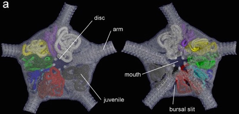
rel="noopener noreferrer"} *Afr J Mar Sci*), by using microCT scanning
the authors were able to visualize in 3D the brooding chambers and
juveniles inside the brittle star, providing an *in situ* view of the
developing young and the position of the bursae inside the adult. The
data ([all hosted](http://dx.doi.org/10.5524/100130) in our GigaDB
repository) includes [videos](https://youtu.be/-jBKtQ0D998) and
projection, reconstruction images and an additional volume file for easy
viewing.
{.alignright
.size-full .wp-image-2219 loading="lazy" decoding="async" width="150"
height="150"}Large-data sharing is an essential part of making research
both reproducible and reusable in the broadest sense. Following our
[\"cyber
centipede\"](http://blogs.biomedcentral.com/gigablog/2013/10/28/the-cyber-centipede-giving-online-species-descriptions-a-leg-up/){target="_blank"
rel="noopener noreferrer"}, [scanned
Earthworms](http://blogs.biomedcentral.com/gigablog/2014/05/19/the-early-earthworm-catches-on-to-full-data-release/){target="_blank"
rel="noopener noreferrer"}, and [MRI\'s of sea
urchins](http://www.gigasciencejournal.com/content/3/1/21){target="_blank"
rel="noopener noreferrer"}, this is our biggest imaging dataset to date,
and further demonstrates the field of morphology is becoming a \"big
data\" field. Following previous [author Q&A\'s in
GigaBlog](http://blogs.biomedcentral.com/gigablog/tag/qa/){target="_blank"
rel="noopener noreferrer"}, here we ask first author Jannes Landschoff
for some insight into why he shared his data via us.
**Can you say a little bit about yourself and the Department of
Biological Sciences and Marine Research Institute at UCT?**
I am currently a PhD student at the University of Cape Town and after my
Masters project on brittle stars I have now moved on to a biodiversity
project on hermit crabs. Born close to the coast in Northern Germany,
marine life has always fascinated me. This was the reason for moving to
Cape Town, where a lot of the marine life, especially marine
invertebrates, are very much unexplored, and where there is still a lot
to discover.
The University of Cape Town is historically strong in marine research
and I am grateful for my training in the [Ma-Re
Institute](http://www.biologicalsciences.uct.ac.za/bio/about/mare){target="_blank"
rel="noopener noreferrer"}, which functions as a link between the
Biological Sciences and Oceanography. For me as a young researcher with
a large interest in marine biodiversity, living and working in a country
like South Africa and studying at UCT is a privilege.
**What attracted you to work on brittle stars and their reproduction in
particular?**
My work on brittle stars started by a coincidence and initially we
wanted to study something completely different. A recreational diver had
sent us pictures of a potential pseudocopulation of two adult brittle
stars. Those pictures of brittle stars mating attracted our interest, in
particular because it seemed as if there was a connection, like a penis
structure between them. The literature and all the experts we consulted
said that this was nonsense, and we pretty soon realised they were
right.
{.wp-smiley
style="height: 1em; max-height: 1em;"} However, by then we had already
come up with more ideas (including the scans), so I stayed with the
group for my masters project and studied the unusual reproduction of a
large brooding brittle star. At that time, nothing was known about that
species although it is very common at our coasts.
\*\*\
Why did you decided to study this organism using micro computed
tomography, and what were you hoping to find using it?\*\*
Our goal was to visualise the very large brooded juveniles inside the
adults without disturbing the surrounding tissues. At first, we purely
wanted to show the feasibility, but we soon realised how amazing the
three-dimensional visualisations looked and that our rotation movies
attracted much interest.
[{.aligncenter
.size-full .wp-image-1617 loading="lazy" decoding="async"
srcset="http://gigasciencejournal.com/blog/wp-content/uploads/2015/11/Figure-2.jpeg 492w, http://gigasciencejournal.com/blog/wp-content/uploads/2015/11/Figure-2-300x143.jpeg 300w"
sizes="(max-width: 492px) 100vw, 492px" width="492"
height="235"}](http://gigasciencejournal.com/blog/wp-content/uploads/2015/11/Figure-2.jpeg)\
**What do you hope others will do this data, and where do you think the
field of morphology is going now we have these techniques?**
Because our focus lay on the brooded juveniles, I believe that our data
have more information than we actually used for our studies. Those
information include the entire morphology of the brittle stars, which we
did not analyse at all. I think that in future, most morphological
studies on marine invertebrate will make use of non-destructive scanning
techniques. Apart from that, I would like to see other studies similar
to ours using the scan data for comparison.
**What do you think is the biggest hurdle for scientists in using these
kinds of advanced imaging techniques? Why have so few echinoderm species
been studied this way so far?**
I think the main constraint of these techniques is not the scanning
itself, but the analysis of the scans which can take a long time and
require a profound knowledge of the analysis computer programs as well
as the right hardware. The scans can be large and require a lot of
computing power as well. In our case, we analysed the data manually,
this meant a long time sitting in front of the PC going through
thousands of image stacks and manually separating the juveniles from the
adult tissue. I was only able to do that because it was my masters
thesis and I had enough time. For studies with a bigger sample size,
automated analyses are needed. Automated processes already exist, but
applying these in practice can often result in different quality for
different species and samples.
**Working in South Africa and off the grid of a lot of the fast
international research networks, have there been any challenges in
working with and trying to disseminate a dataset such as this that is in
the 100GB range? As this type of data sharing isn\'t currently mandatory
what drove you to go to the effort?**
Working in a country like South Africa, we are used to problems that
many people in the western world may hardly ever think of. For example,
uploading a dataset of 100GB takes a long time, especially with slow
internet connections. When the upload is hit by a sudden power outage,
it might happen that you have to start the upload again. In fact, it
took us several months to submit the data! I still think it is worth it
because it sharing the data makes my research more transparent and more
accessible.
**Now you have managed to visualise and compare brooding at this level
in the species, what is next for your research in this area? Are you
working on any other species in the same manner?**
As I have mentioned above I have now moved on to crustacean research,
but scanning techniques can be equally valuable in crustacean biology.
Although I am not planning any scanning of crustaceans right now, I am
grateful to have the skills and facilities to do that sort of work.
Apart from my PhD project, we are still working on a few other brooding
species of echinoderms. As those project can teach you a lot about
internal anatomy, are hands on, and result in fascinating images, they
make great study opportunities for undergrad students. Next year, we
will be publishing another reproduction study on a coastal brittle star,
*Amphiura capensis*, of which one scan is already included in the
published data set.\
\*\*\
\*\*
### **References**
1\. Landschoff J, Du Plessis A, Griffiths CL. 2015. A dataset describing
brooding in three species of South African brittle stars, comprising
seven high-resolution, X-ray micro tomography scans. *GigaScience* 2015:
4:52.
doi:[10.1186/s13742-015-0093-2](http://dx.doi.org/10.1186/s13742-015-0093-2){target="_blank"
rel="noopener noreferrer"}.
2\. Landschoff, J; Du Plessis, A; Griffiths, C, L (2015): 3-dimensional
microCT reconstructions of brooding brittle stars GigaScience Database.
[http://dx.doi.org/10.5524/100130](http://dx.doi.org/10.5524/100130){target="_blank"
rel="noopener noreferrer"}
3\. Landschoff, J. and Griffiths, C.L. (2015) 3D visualisation of
brooding behaviour in two distantly-related brittle stars from South
African waters. Afr J Mar Sci; 37(4).
doi:[10.2989/1814232X.2015.1095801](http://dx.doi.org/10.2989/1814232X.2015.1095801){target="_blank"
rel="noopener noreferrer"}.
The post [Brooding Brittle Star Babies. Q&A with Jannes
Landschoff](http://gigasciencejournal.com/blog/brooding-brittle-star-babies-qa-jannes-landschoff/){rel="nofollow"}
appeared first on
[GigaBlog](http://gigasciencejournal.com/blog){rel="nofollow"}.